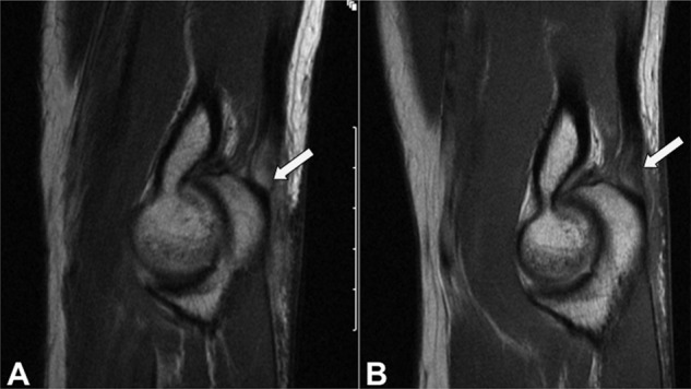
Figure 5.

Sagittal proton density MRI sequence (A) demonstrates an acute partial tear of the triceps insertion (arrow) superimposed on preexisting tendinosis. One year later (B), a repeat MRI shows improved appearance of the triceps tendon but residual tendinosis (arrow). Images courtesy of Hollis G. Potter, MD.
