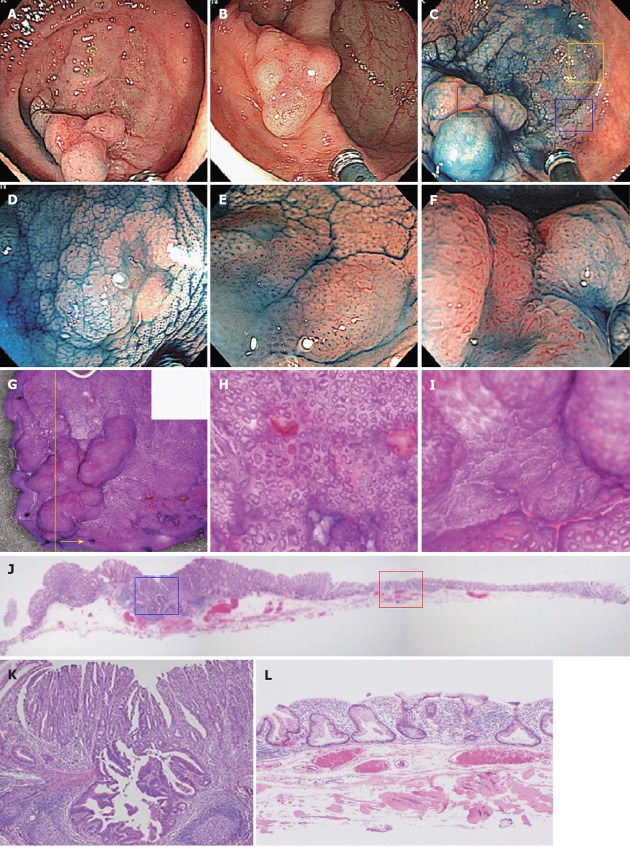
Figure 3.
Adenocarcinoma (tub1) in sessile serrated adenomas/polyps case. A 60-year-old woman had a tumor lesion in the cecum, which was flat and elevated, that is a so-called lateral spreading tumor (LST), 45 mm in diameter. A, B: Standard view. A 45-mm LST lesion with large amounts of mucin is evident in the cecum. The flat portion is somewhat discolored compared with the surrounding mucosa. The protruded portion on the anal side is reddish in the center; C: Indigo carmine staining. The center of the protruded portion on the anal side is flat; D: Indigo carmine staining, enlarged image (yellow square in 3C). Type II-Long pit pattern is evident; E: Indigo carmine staining, enlarged image (blue square in 3C). Type II-Open pit pattern is evident; F: Indigo carmine staining, enlarged image (black square in 3C). We diagnosed as type VI pit pattern, because high density of crypts and irregular pit pattern were evident; G: Comparison of stereomicroscopic and colonoscopic images; H: Enlarged image of flat portion. Although basically type II, dilated duct openings are evident; I: Enlarged image of protruded portion on the anal side. An irregular surface architecture is evident; J: HE magnifying glass image (yellow line in Figure 3G). We examined HE staining (Figure 3J-L) using the yellow cutting line of endoscopically resected tissue (yellow arrow side tissue); K: Central part of the protrusion on the anal side (blue square in Figure 3J). Highly differentiated ductal cancer corresponding to type VI pits is evident; L: Dilatation of crypts and deformation in the horizontal direction at the bottom of the crypts are evident in the flat portion (red square in Figure 3J). Sessile serrated adenomas/polyps was diagnosed.

