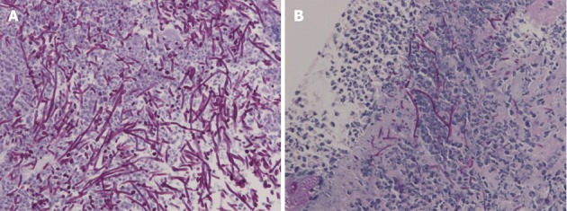
Figure 2.

Biopsy demonstrated no malignancy but numerous hyphae. A: Light micrograph of the specimen biopsied from the original ulcer demonstrating hyphae in the ulcer slough [Periodic acid-Schiff (PAS)/diastase, original magnification × 400]; B: Biopsy from the recurrent ulcer illustrating hyphae of Candida on the ulcer edge (PAS/diastase, original magnification × 400).
