Figure 3.

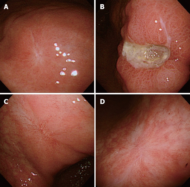
Endoscopic photographs. A: The white scar of the original ulcer; B: The recurrent ulcer on the lesser curvature of the lower gastric body; C: The red scar of the recurrent ulcer; D: The transition from the red to white scar in 3 mo.

Endoscopic photographs. A: The white scar of the original ulcer; B: The recurrent ulcer on the lesser curvature of the lower gastric body; C: The red scar of the recurrent ulcer; D: The transition from the red to white scar in 3 mo.