Fig. 3.
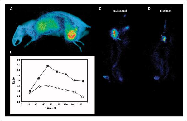
Whole-body planar scintigraphy of Dunning prostate R3227-AT1tumor-bearing rats. A, rats bearing ~20 mm diameter tumors were injected i.v. with 5 MBq of [74As]bavituximab. The rats were imaged on a phosphor plate at various time points after injection. Representative image 72 h after injection. The image is overlaid on an X-ray picture to provide anatomic correlation. B, ratio of uptake of [74As]bavituximab in tumor versus upper organs (liver, lung, heart) at various time points after injection. ●, outer tumor regions; ◯, entire tumor. At 24 h after injection, no obvious contrast was observed, but at 48 h, the tumor became clearly visible and by 72 h, the tumor-to-organ ratio was the highest. C–D, scintigraphy of rats injected with 3 MBq [77As]bavituximab or [77As]rituximab (negative control). Images acquired with 30 min of exposure time at 72 h. Eight-fold higher uptake of bavituximab than of the control antibody was observed in the tumor.

