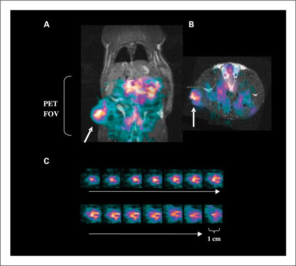
Fig. 5.
Small animal PET. A and B, small animal PET images obtained from a Dunning prostate R3227-AT1 tumor-bearing rat 48 h after injection of 10 MBq of [74As]bavituximab coronal (A) and transaxial (B). PET intensity is overlaid on slices obtained by three-dimensional MRI. [74As]bavituximab localized to the tumor (arrows) and was also visible in the blood pool of normal organs.The PET field of view (FOV) is indicated by the bracket. C, images of 1-mm sequential tumor slices from the three-dimensional data sets.

