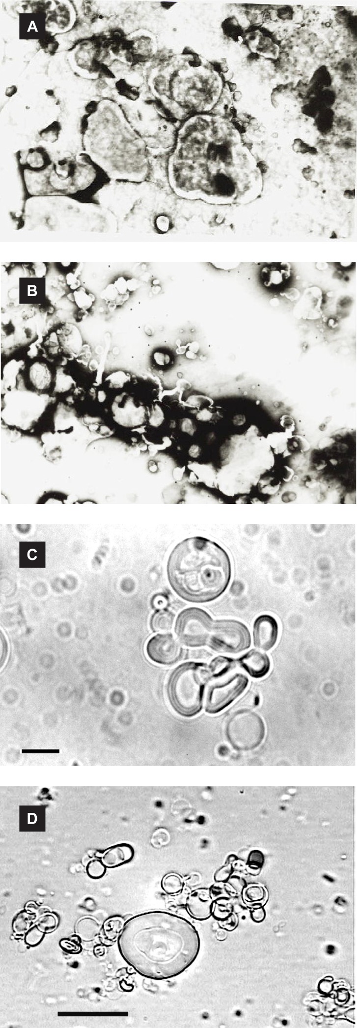
Figure 1.
Micrographs (×1000 magnification) of insulin-loaded niosomes (7:3 molar ratio of surfactant/cholesterol) prepared by classic film method: negative staining transmission electron microscopy micrographs of (a) Brij 52 vesicles, (b) Span 60 niosomes, and optical microscopy pictures of (c) Span 60 vesicles, (d) Brij 92 niosomes; Bar = 10 µm.

