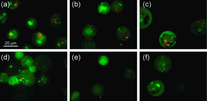
Figure 9.

Acridine orange staining of GLC4 cell line after exposure of 1 h to different receptors: (a) untreated cells (control), (b) cells treated with receptor 2, (c) cells treated with receptor 3, (d) cells treated with receptor 7, (e) cells treated with receptor 8, (f) cells treated with receptor 9. (a–c) Cells with granular orange fluorescence in the cytoplasm; (d–f) cells with complete disappearance of orange fluorescence cytoplasm granules.
