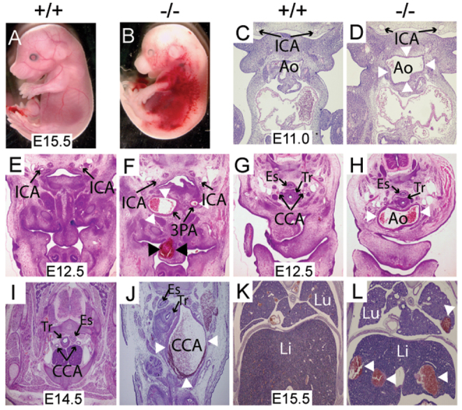
Fig. 1.

Mkl2–/– embryos exhibit hemorrhage and aneurysmal dilation of the great arteries. (A,B) E15.5 wild-type (+/+) and Mkl2–/– (–/–) embryos demonstrating diffuse hemorrhage in the E15.5 mutant embryo. (C,D) Hematoxylin and Eosin-stained transverse sections of E11.0 wild-type and Mkl2–/– embryo, demonstrating dilated aortic sac (Ao) (arrowheads) in the mutant embryo. Original magnification was ×20. ICA, internal carotid artery. (E,F) Hematoxylin and Eosin-stained transverse section of E12.5 wild-type and Mkl2–/– embryo, demonstrating dilation of the 3rd PA artery and dilation and hemorrhage (black arrowheads) of the lingual vessels in the Mkl2–/– mutant embryo. Original magnification was ×20. White arrowheads indicate aneurysm. (G,H) Hematoxylin and Eosin-stained section of E12.5 wild-type and Mkl2–/– embryo, showing dilation of the mutant aorta (Ao, white arrowheads) extending to the level of the common carotid artery (CCA). Original magnification was ×20. ES, esophagus; Tr, trachea. (I,J) Hematoxylin and Eosin-stained transverse section of E14.5 wild-type and Mkl2–/– embryo, demonstrating aneurysmal dilation of the mutant common carotid artery (CCA). Original magnification was ×20. Arrowheads indicate aneurysm. (K,L) Hematoxylin and Eosin-stained frontal sections of E15.5 wild-type and Mkl2–/– embryos showing intra-parenchymal hemorrhages (white arrowheads) in the liver (Li) and lung (Lu) of the mutant embryo. Original magnification was ×20.
