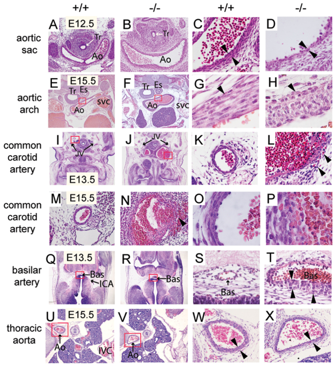
Fig. 2.

Disruption of the tunica media and aneurysm formation in Mkl2–/– embryos. (A-D) Hematoxylin and Eosin-stained transverse section of E12.5 wild-type (+/+) and Mkl2–/– (–/–) embryos, demonstrating dilation of the mutant aortic sac in the Mkl2–/– mutant aorta (Ao). Tr, trachea. (C,D) High-power magnification of the wall of the aortic sac reveals thinning of the tunica media to a single layer of cells (arrowheads) in some segments of the Mkl2–/– mutant aortic sac. Original magnifications were ×100 (A,B) and ×400 (C,D). Tr, trachea. (E-H) Hematoxylin and Eosin-stained transverse section of the aortic arch (Ao) of E15.5 wild-type and Mkl2–/– embryo, demonstrating dilation of mutant aorta arch (Ao). The red rectangles in E and F indicate the locations of G and H. (G,H) High-power magnification of the tunica media (arrowhead) of the control artery reveals spindle-shaped SMCs oriented circumferentially (G); by contrast, medial SMCs populating the mutant artery appear polygonal lacking predominant orientation with obvious gaps between cells (H). Original magnifications were ×40 (E,F) and ×1000 (G,H). (I-L) Hematoxylin and Eosin-stained transverse section demonstrating aneurysmal dilation of the common carotid arteries of E13.5 wild-type and Mkl2–/– embryos. The location of the jugular vein (JV) is indicated. The red rectangles indicate the locations of K and L. (K,L) High-power magnification reveals thinning of the tunica media and rupture through the arterial wall (arrowheads) of the in the Mkl2–/– embryo (L). By contrast, spindle-like SMCs compose the media of the control artery (K). Original magnifications were ×20 (I,J) and ×200 (K,L). (M-P) Hematoxylin and Eosin-stained section showing the common carotid artery of E15.5 wild-type and Mkl2–/– embryo, demonstrating aneurysmal dilation, dissection and rupture (arrowhead) in the Mkl2–/– embryo (N). (O,P) High-power magnification demonstrates alignment of spindle-shaped SMCs surrounding the lumen of the control carotid artery (O). By contrast, SMCs of the Mkl2–/– artery are polygonal, lacking predominant orientation with intramural hemorrhage observed between cells (P). Original magnifications were ×100 (M,N) and ×1000 (O,P). (Q-T) Hematoxylin and Eosin-stained section of the basilar artery (Bas) of E13.5 wild-type and Mkl2–/– embryos demonstrating dilation of the mutant basilar artery (Bas) (R,T). The red rectangles in Q and R indicate the locations of S and T. (S,T) High-power magnification reveals disorganization of the tunica media (arrowheads) in the Mkl2–/– artery compared with the control basilar artery, which comprises a single layer of SMCs. Original magnifications were ×40 (Q,R) and ×400 (S,T). ICA, internal carotid artery. (U-X) Hematoxylin and Eosin-stained section showing the descending thoracic aorta of E15.5 wild-type (U,W) and Mkl2–/– (V,X) embryos. The red rectangles in U and V indicate the locations of W and X. Slight thinning of the tunica media (arrowheads) is observed in the Mkl2–/– mutant artery (X) compared with the control aorta (W). Original magnifications were ×40 (U,V) and ×200 (W,X). IVC, inferior vena cava. See also supplementary material Fig. S2.
