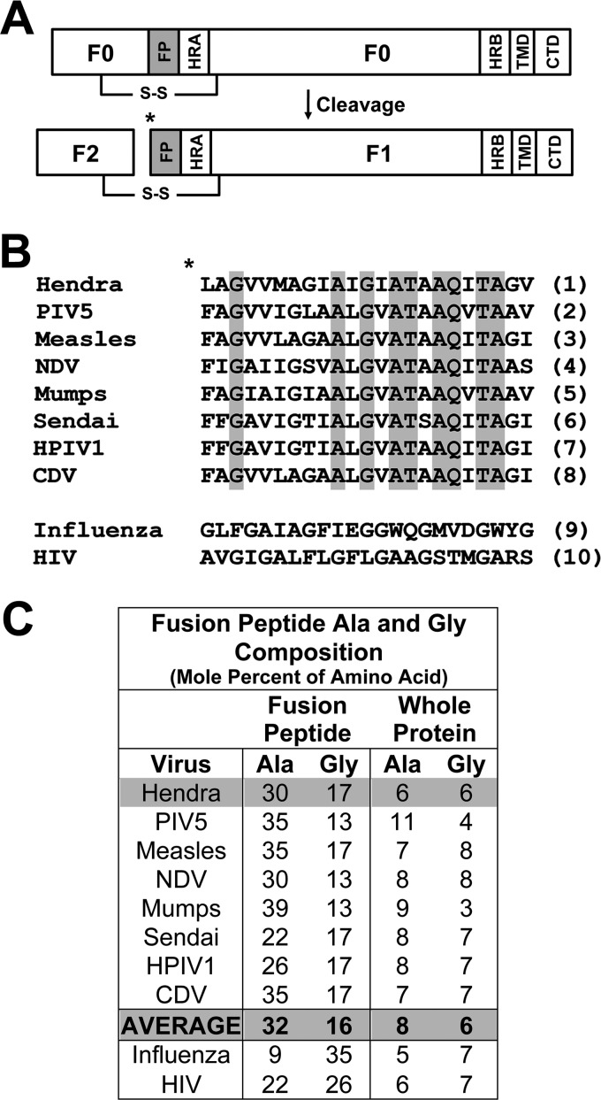
FIGURE 1.
Amino acid composition of fusion peptides from class I fusion proteins. A, shown is a diagram of a paramyxovirus F protein in the uncleaved (F0) and cleaved, fusogenically active F1+F2 forms. HR, heptad repeat; CTD, C-terminal domain (C-tail); * denotes site of proteolytic cleavage. B, shown is alignment of paramyxovirus fusion peptides with completely conserved amino acids shaded and compared with fusion peptides from influenza HA and HIV Env. References for sequences are as follows: Hendra (5), PIV5 (59), measles (60), NDV (61), Mumps (62), Sendai (63), HPIV1 (human parainfluenza virus type 1 (64)), CDV (canine distemper virus (65)), influenza (66), HIV (22). C, percent alanine and glycine composition of the fusion peptides is shown in B as compared with the whole protein.

