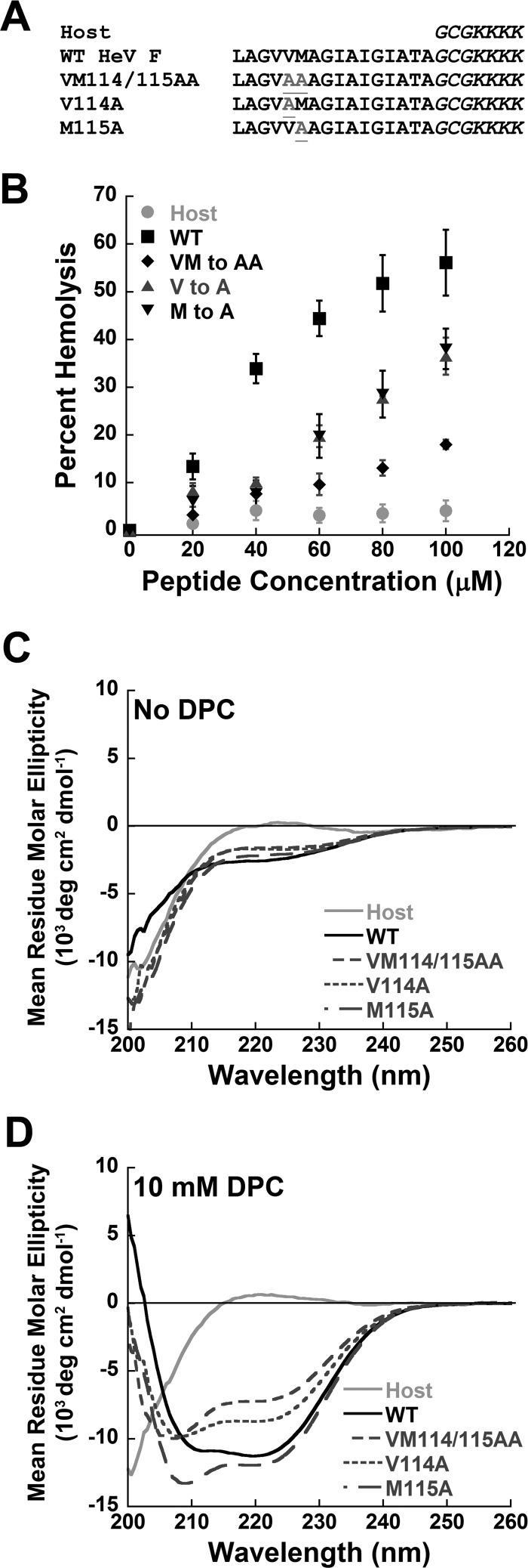
FIGURE 4.
Hemolytic ability and CD spectra of synthetic fusion peptides. A, shown are synthesized peptides where the first 16 amino acids of the FP for either wild-type or mutant Hendra F are coupled to a flexible highly charged host peptide. B, hemolytic ability of synthetic fusion peptides is shown. Chicken RBCs were incubated with increasing concentrations of peptide for 45 min at 37 °C. Supernatants were then cleared by centrifugation at 11,000 × g, and absorbance was measured at 520 nm. All values are expressed as a percent of maximum hemolysis determined by RBC lysis with 0.5% Triton X-100. Data are presented as the means ± S.E. for at least three independent experiments. C, CD spectra of all five peptides in the absence of DPC in 5 mm Hepes, 10 mm MES pH 7.4 with peptide concentrations of 100 μm are shown. D, shown are spectra in the presence of 10 mm DPC micelles prepared in the above buffer as described under “Experimental Procedures.” Spectra in both C and D are representative of one of three independent experiments where each spectrum is an average of four scans.

