FIGURE 1.
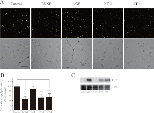
Neurotrophic factors reduce cell surface neprilysin activity in primary cortical/hippocampal neurons. A, primary cortical/hippocampal neurons infected with SFV-hNEP were incubated with BDNF (100 ng/ml), NGF (100 ng/ml), NT-3 (100 ng/ml), or NT-4 (100 ng/ml) for 24 h, after which they were subjected to the neprilysin activity-staining assay. The top panels show fluorescence images representing neprilysin activity, and the bottom panels show phase-contrast images merged with the top panels. Scale bar, 100 μm. B, quantification of the fluorescence signal areas, indicated as average ± S.D. (error bars) (n = 5). ***, p < 0.01 compared with control. C, primary neurons were incubated with BDNF (100 ng/ml), NGF (100 ng/ml), NT-3 (100 ng/ml), or NT-4 (100 ng/ml) for 30 min and then subjected to Western blot analysis to measure the phosphorylation level of the neurotrophic factor receptor, Trk, using antibodies against Trk receptor (bottom) and phosphorylated Trk (p-Trk; top). At least three independent experiments were repeated.

