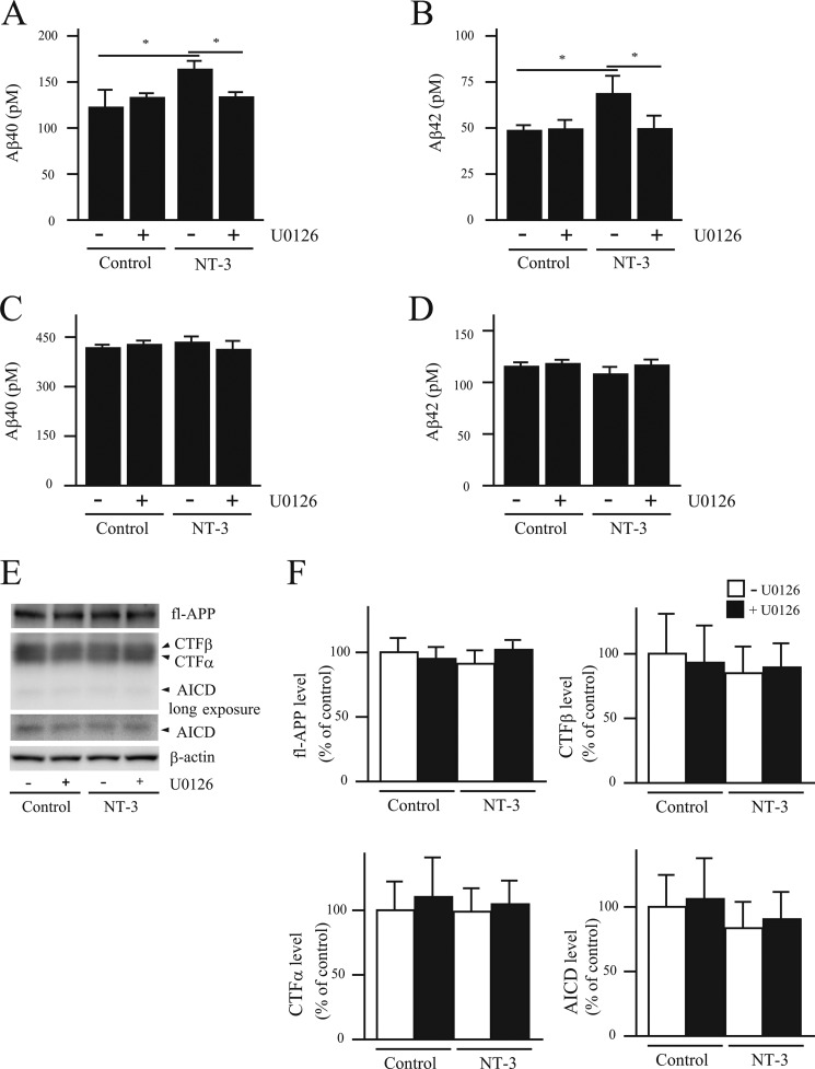
FIGURE 3.
NT-3 induces increased Aβ levels via the MEK/ERK pathway. A–D, the culture medium of primary neurons derived from wild-type (A and B) or NEP-deficient mouse embryos (C and D) was collected 24 h after NT-3 (100 ng/ml) treatment with or without U0126 (1 μm) and then subjected to Aβ ELISA. Aβ40 (A and C) and Aβ42 (B and D) levels were measured using an Aβ-ELISA kit. Each column with error bar represents the mean ± S.D. (n = 4). *, p < 0.05 compared with control and co-treatment. E and F, the effect of neurotrophic factors on Aβ generation was investigated by measuring the levels of full-length APP (fl-APP), CTFα, CTFβ, APP intracellular domain (AICD), and β-actin in the primary neurons by Western blot. Intensities of each band were quantified by densitometric analysis, and data represent the mean ± S.D. (n = 5).

