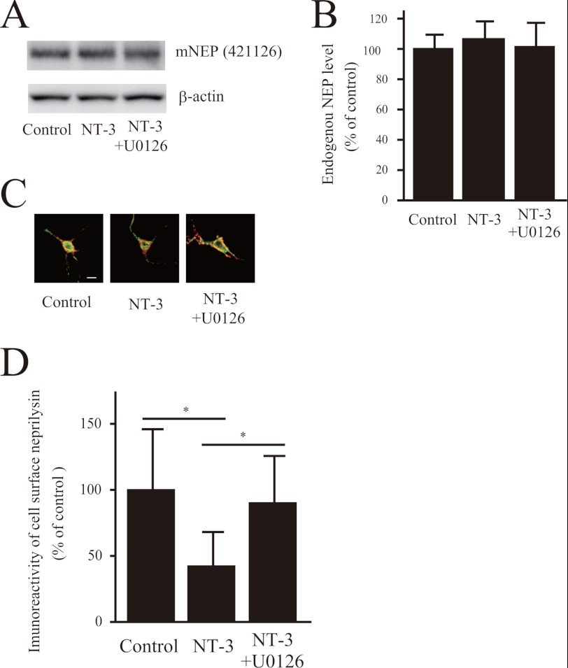
FIGURE 4.
NT-3 regulates neprilysin localization via the MEK/ERK pathway. A and B, the effect of NT-3 on expression levels of neprilysin in primary neurons was analyzed by Western blot analysis. The experiments were repeated four times, and the results are presented as mean values ± S.D. (error bars). C and D, double staining for neprilysin and biotinylated proteins located on the cell surface. Primary cortical neurons infected with SFV-hNEP were treated with NT-3 (100 ng/ml) for 24 h. The cell surface proteins were subsequently cross-linked with biotin, after which the cells were double-stained with neprilysin antibody (56C6; green) and Alexa 546-conjugated streptavidin (red). The images of the green and the red signals were merged, yellow representing cell surface neprilysin. Scale bar, 50 μm. The ratio of cell surface neprilysin levels was quantified by image analysis. Data represent the mean ± S.D. (n = 15). *, p < 0.05 compared with control and co-treatment.

