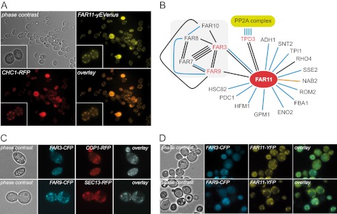
FIGURE 3.
Subcellular localization of Far proteins. A, subcellular localization of the Far11-yEVenus and Chc1-RFP fusion proteins. A magnification of two cells is shown in the inset. B, schematic representation of the physical interaction network of Far11 according to the information deposited at SGD. The number of lines indicates the number of experiments reported and the color of the lines indicates the type of experiment described: black lines represent two-hybrid analyses; blue lines indicate affinity-capture-MS experiments; and the red line corresponds to an affinity-capture RNA experiment. C, subcellular localization of: Far3-CFP and Cop1-RFP (upper panels); Far9-CFP and Sec13-RFP (lower panels). D, colocalization of Far11-yEVenus with either Far3-CFP (upper panels) or Far9-CFP (lower panels).

