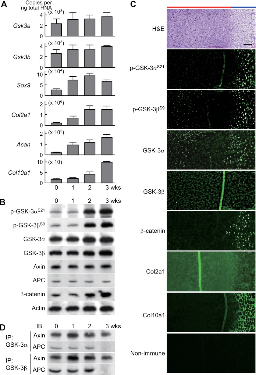
FIGURE 1.
A, mRNA levels of Gsk3a, Gsk3b, and early (Sox9, Col2a1, and Acan) and later (Col10a1) chondrocyte differentiation markers during culture of mouse chondrogenic ATDC5 cells in differentiation medium (ITS) for 3 weeks. Data are expressed as means ± S.D. for three wells/group. B, immunoblotting using antibodies to Ser-21-phosphorylated GSK-3α (p-GSK-3αS21) and Ser-9-phosphorylated GSK-3β (p-GSK-3βS9), GSK-3α, GSK-3β, axin, APC, and β-catenin with actin as the loading control during differentiation of cultured ATDC5 cells as above. C, H&E and immunofluorescence with antibodies to the indicated proteins, as well as the nonimmune control, in the proximal tibias of mouse embryos (E18.5). Scale bar, 100 μm. Red and blue bars indicate layers of proliferative and hypertrophic zones, respectively. D, physical association of GSK-3 with scaffolding proteins axin and APC by immunoprecipitation (IP) and immunoblotting (IB) analyses during differentiation of cultured ATDC5 cells as above.

