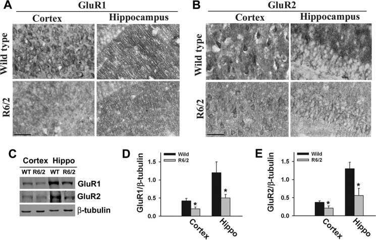
FIGURE 6.
Expression of GluR1 and GluR2 is decreased in R6/2 mouse brain. A and B, representative immunohistochemical staining of GluR1 and GluR2 in the cortices and hippocampuses of wild-type and R6/2 mice. Scale bars = 10 μm. C, immunoblot analysis of GluR1 and GluR2 in the cortex and hippocampal (Hippo) regions of wild-type and R6/2 mouse brains (10–12 weeks old). D and E, GluR1 and GluR2 blots obtained from four different mice in each group were quantitated using NIH ImageJ analysis software and normalized against β-tubulin. *, p < 0.05 in comparison with wild-type mice.

