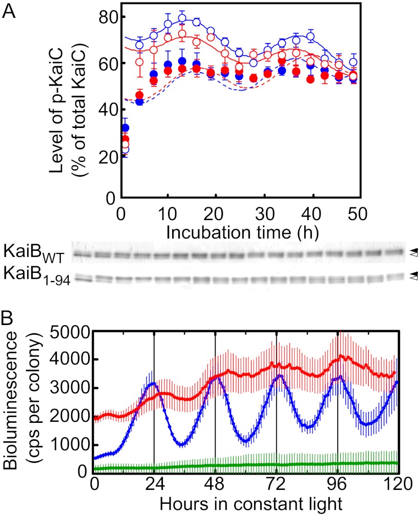
FIGURE 3.
Circadian oscillations in vitro and in vivo. A, shown are circadian oscillations in vitro. Generation of temperature-compensated circadian oscillations in vitro in the level of p-KaiC by KaiB1–94 in an in vitro KaiABC clock system is shown. Reaction mixtures containing KaiA, KaiBWT or KaiB1–94, and KaiCWT (0.5 μm each) were incubated in 20 mm Tris-HCl buffer (pH 7.5) containing 1 mm ATP, 5 mm MgCl2, and 150 mm NaCl at 25 °C or 40 °C for the periods indicated and were subjected to SDS-PAGE on 12.5% gels. The gels were stained with CBB, and the relative amounts of np-KaiC and p-KaiC were estimated by densitometry. Typical photographs from three independent experiments are shown. Circadian oscillations in the level of p-KaiC were analyzed by RAP, and the simulated curves are shown. Values shown are the means ± S.D. from triplicate measurements. KaiBs added: open red circles, KaiB1–94 (40 °C); open blue circles, KaiBWT (40 °C); closed red circles, KaiB1–94 (25 °C); closed blue circles, KaiBWT (25 °C). Closed arrows, p-KaiC; open arrows, np-KaiC. B, shown are circadian bioluminescence rhythms of a T. elongatus PpsbA1::Xl luxAB reporter strain carrying kaiBWT or kaiB1–94 in a kaiB-null genetic background. Bioluminescence rhythms of T. elongatus PpsbA1::Xl luxAB reporter strains carrying kaiBWT (WT, blue, n = 10), kaiB1–94 (KaiB1–94, red, n = 13), and no additional kaiB gene (KaiB-null, green, n = 75) in a kaiB-null background (the kaiB gene had a nonsense codon just downstream of its initiation codon) were measured at 41 °C. Data points and error bars indicate the mean ± S.D. from n independent samples. cps, counts per s.

