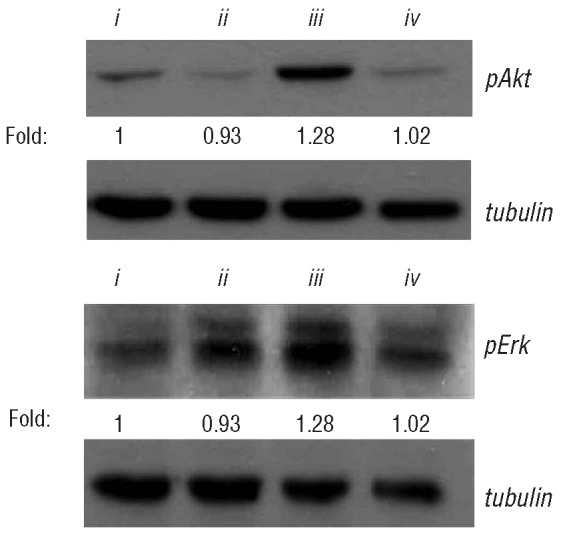
Figure 4.

Western blot assays for pAkt and pErk in Jurkat T cells stimulated with anti-CD3 plus anti-CD28 (i) untreated, or treated with (ii) sirolimus 5nM, (iii) bortezomib 100 nM and (iv) sirolimus 5 nM plus bortezomib 100 nM. The numbers shown below the bands indicate the quantitative measurement of the fold change with respect to the untreated sample; data from 4 experiments are shown.
