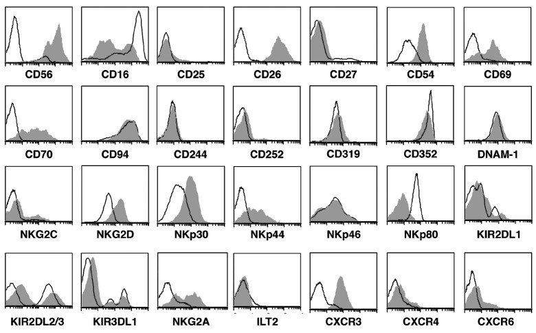
Figure 1.
Exp-NK cells have an activated phenotype. Flow cytometry confirms the increased cell surface density of NKG2D, NKp30, NKp44, CD26, CD56, CD54, CD69, CD70, and the chemokine receptor CXCR3. Open peaks represent non-exp-NK, shaded peaks represent exp-NK. One representative result from 12 experiments is shown.

