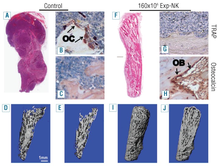
Figure 4.
Exp-NK cell treatment protects bone from myeloma-induced osteolysis. On day 29, at the termination of the experiment depicted in Figure 3, implanted fetal bones were excised and subjected to immunohistochemistry and micro-computed tomography (CT). H&E, TRAP, and osteocalcin staining revealed gross loss of bone, increased numbers of osteoclasts, and reduced numbers of osteoblasts in the control group (A–C) compared to those in the mice treated with 160×106 exp-NK cells (F–H). Micro-CT further confirmed the dramatic difference in implanted human bone structure between control and exp-NK cell–treated groups. Three-dimensional reconstructions of the bones are shown (D, I), as well as longitudinal sections through the midpoint from each specimen (E, J). Heavy bone resorption is clearly seen in the control group (D, E), while bone resorption is not prominent in the exp-NK cell–treated group (I, J).

