FIGURE 2.
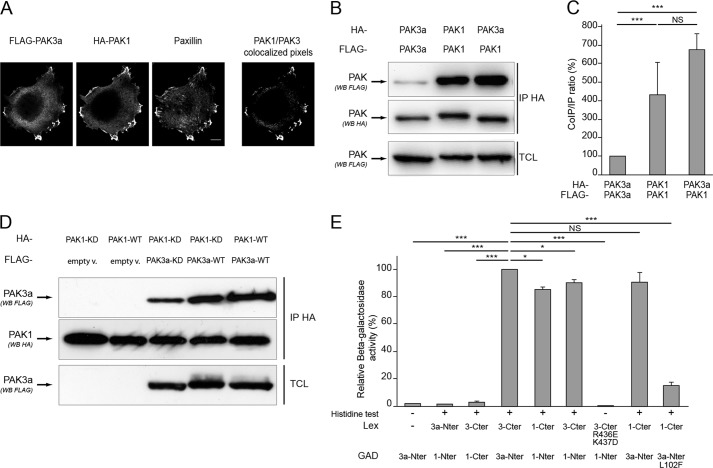
PAK3a forms heterodimers with PAK1. A, PAK1 and PAK3 colocalize at focal adhesions of HeLa cells. FLAG-PAK3a-KD and HA-PAK1-KD plasmids were transfected in HeLa cells. Immunolabeling was performed using FLAG and HA antibodies to detect PAK proteins and paxillin antibodies to identify focal adhesions. The PAK1/PAK3 colocalized pixels are shown (right). B, PAK3a co-immunoprecipitates with PAK1. HA- and FLAG-tagged PAK3a-KD and PAK1-KD plasmids were co-transfected in HeLa cells then cell lysates were HA-precipitated (IP HA). The co-immunoprecipitated PAK proteins were revealed by anti-FLAG Western blotting (first panel). HA-precipitated proteins and TCL were controlled using the corresponding antibodies as indicated (second and third panels, respectively). Images shown are representative of three independent experiments. C, amount of FLAG-PAK proteins co-immunoprecipitated compared with the amount of HA-precipitated PAK proteins. Results are expressed relative to the PAK3a/PAK3a interaction. Comparison with Student's t test: NS, p > 0.05; ***, p < 0.001, n = 3. D, wild-type PAK isoforms interact together. Cells were co-transfected with HA-PAK1-KD and FLAG-PAK3a-KD (third lane), HA-PAK1-KD, and FLAG-PAK3a-WT (fourth lane), and HA-PAK1-WT and FLAG-PAK3a-WT (fifth lane). HA-proteins were immunoprecipitated (second panel) and co-immunoprecipitated FLAG proteins were visualized by anti-FLAG Western blotting (first panel). Co-transfection of PAK1 with empty vector (empty v.) was done as control (first two lanes). Images shown are representative of three independent experiments. E, analysis of the PAK1/PAK3a interaction using the two-hybrid assay. Yeasts growth on histidine minus media indicated a protein-protein interaction (+). β-Galactosidase activity was expressed relative to the PAK3a-Nter/PAK3a-Cter interaction. Comparison with Student's t test: NS, p > 0.05; *, p < 0.05; ***, p < 0.001; n = 3.

