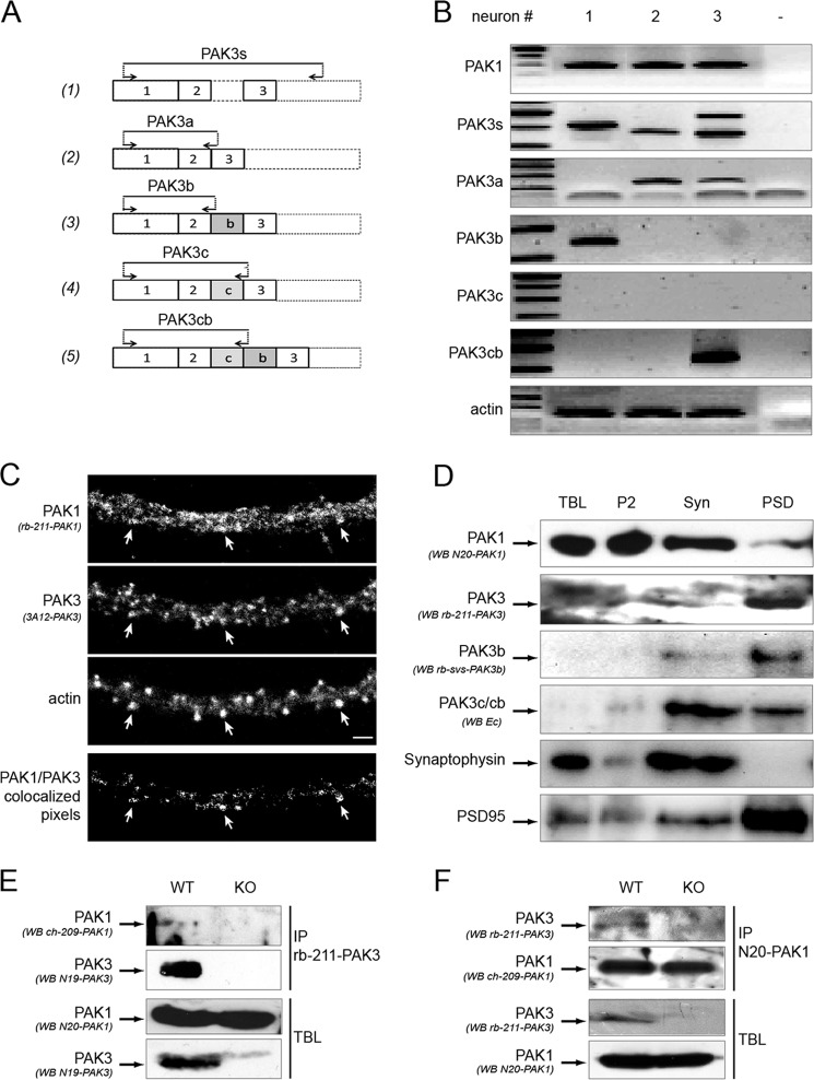
FIGURE 5.
PAK3 and PAK1 are coexpressed in single neurons, colocalize in dendritic spines, co-purified with the postsynaptic density, and co-immunoprecipitate in brain extracts. A, schematic representation of the location of the different PAK3 oligonucleotides used for single-cell RT-PCR. Forward and reverse oligonucleotides are indicated by arrows. Primers were chosen near or within the exons 2–3 portion to detect all the PAK3 cDNAs (1), and at exon/exon junction to specifically detect PAK3a (2), PAK3b (3), PAK3c (4), and PAK3cb (5) cDNAs. B, PAK3 and PAK1 are co-expressed in single neuron. RNAs from single pyramidal neurons were extracted and RT-PCR was performed using the different primer sets. Actin amplification was used as control (panel 7). Specific primer sets were used for PAK1 (first panel), all PAK3s (second panel), PAK3a, b, c, cb (third to sixth panel). C, PAK3 and PAK1 partially colocalize in dendritic spines of differentiated hippocampal neurons. Endogenous PAK proteins were immunolabeled with rb-211-PAK1 antibodies (first panel) and monoclonal 3A12 PAK3 antibodies (second panel). Dendritic spines were visualized by Alexa-633-phalloidin labeling (third panel) on dissociated hippocampal neurons at DIV21. On the PAK1/PAK3 colocalization image (fourth panel), white arrows indicate some spines where PAK1 and PAK3 proteins are co-expressed. Scale bar, 2 μm. D, PAK3 and PAK1 partially co-purify with some synaptosomal fractions as shown by Western blots of the different fractions purified from brain lysates. TBL from adult mice brain were fractionated to obtain the second pellet (P2) fraction, the synaptosomal fraction (Syn) and the postsynaptic densities (PSD) fraction. The same amount of protein was loaded for each fraction. The different fractions were probed with either the N20-PAK1 antibodies specific for PAK1 (first panel), the rb-211-PAK3 antibodies that recognized the PAK3s proteins (second panel), the rb-svs-PAK3b antibodies specific for PAK3b variant (third panel), the Ec antibodies that recognized the PAK3c and PAK3cb variants (fourth panel). Western blots with the synaptophysin antibodies as a presynaptic marker (fifth panel), and the PSD-95 antibodies as a postsynaptic density marker (sixth panel) confirm quality of tissue fractionation. Images shown are representative of three experiments. E, PAK1 co-immunoprecipitates with PAK3. TBL were prepared from adult wild-type mice (WT) and as a negative control, from pak3− mice (KO). In TBL, PAK1, and PAK3 proteins were detected by immunoblotting using the N20-PAK1 antibodies (third panel) and the N19-PAK3 (fourth panel), respectively. PAK3 proteins were immunoprecipitated with the N19-PAK3 sera (second panel) and co-immunoprecipitated PAK1 proteins were detected with ch-209-PAK1 (first panel). F, PAK3 co-immunoprecipitates with PAK1. TBL were processed as for E, to analyze PAK3 (third panel) and PAK1 (fourth panel) expression. PAK1 proteins were immunoprecipitated using the N20-PAK1 antibodies (second panel) and co-immunoprecipitated PAK3 proteins were then detected with the rb-211-PAK3 sera (first panel). Images shown are representative of two experiments.

