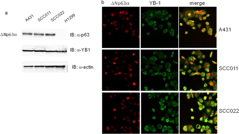
FIGURE 1.
ΔNp63α and YB-1 are co-expressed in squamous carcinoma cell lines. a, whole cell lysates were obtained from H1299 (non-small cell lung carcinoma), A431 (epidermoid carcinoma cell line), and SCC011 and SCC022 (keratinocyte-derived SCC) cells. 30 μg of total protein extracts were separated by SDS-PAGE and subjected to immunoblot (IB). Proteins were detected with specific antibodies as indicated. Images were acquired with CHEMIDOC (Bio-Rad) and analyzed with the Quantity-ONE software. b, A431, SCC011, and SCC022 cells were seeded (2.5 × 10⁵) on a 35-mm dish and grown on micro cover glasses (BDH). After 24 h at seeding, cells were fixed and subjected to double immunofluorescence using rabbit primary YB-1 antibody and Fitch-conjugated secondary antibodies (green). p63 protein was detected using mouse anti-p63 and Cy3-conjugated secondary antibodies (red). Images of merge (yellow) show the co-expression of two proteins.

