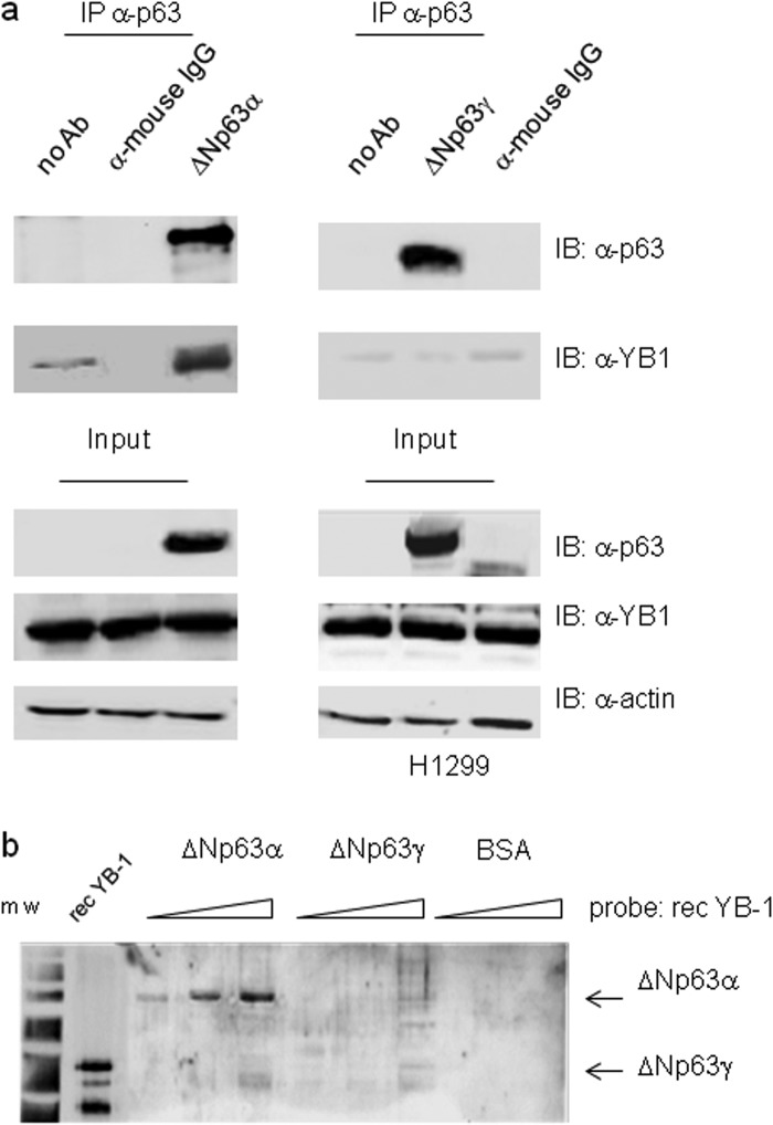
FIGURE 3.
ΔNp63α but not ΔNp63γ interacts with YB-1. a, H1299 cells were transiently transfected with 5 μg of ΔNp63α (left panel) or ΔNp63γ (right panel) expression vectors. Equal amounts (1 mg) of extracts were immunoprecipitated (IP) with anti-p63 antibodies (4A4) or unrelated α-mouse IgG. The immunocomplexes were blotted and probed with anti-p63 and anti-YB-1 antibodies, as indicated. b, far Western analysis. Increasing amounts of purified recombinant ΔNp63α (0.2, 0.5, and 1.5), ΔNp63γ (0.2, 0.5, and 1.5), or BSA (0.5 and 1.5) were subjected to SDS-PAGE. After Coomassie staining to monitor equal loading, the proteins were transferred to a PVDF membrane, and the filter was incubated with purified YB-1 recombinant protein (0.8 μg/ml). After extensive washing, the membrane was subjected to immunoblotting (IB) with YB-1 antibodies followed by ECL detection. Recombinant YB-1 protein (0.1 μg) was used as positive control. m.w., molecular weight markers.

