FIGURE 5.
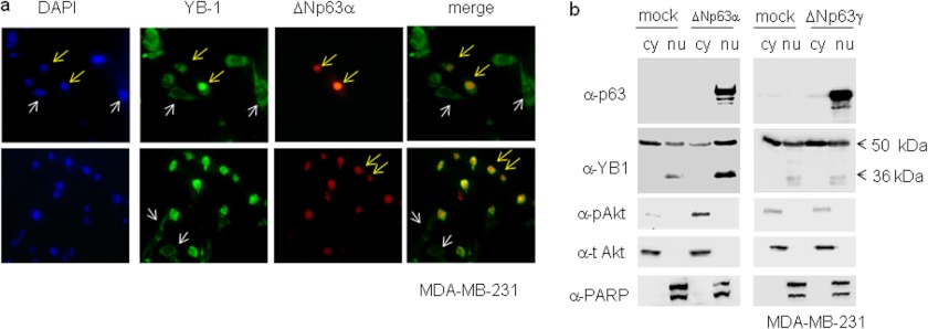
Endogenous YB-1 accumulates in the nucleus of breast cancer cells expressing ΔNp63α. a, MDA-MB-231 cells were seeded at 60% confluency (2.3 × 10⁵) on 24 × 24-mm sterile coverglasses placed in 60-mm dishes and transiently transfected with 1 μg of ΔNp63α expression vector. Cells were fixed and subjected to double indirect immunofluorescence using rabbit primary YB-1 antibody and Fitch-conjugated secondary antibodies (green). p63 protein was detected using mouse anti-p63 and Cy3-conjugated secondary antibodies (red). DAPI was used to stain nuclei (blue). A representative image is given of a cell expressing ΔNp63α and showing nuclear endogenous YB-1 (yellow arrows). A representative image is given showing YB-1 cytoplasmic localization in cells bearing no detectable ΔNp63α expression (white arrows). b, MB-MDA-231 cells were transiently transfected with a fixed amount (5 μg) of an empty vector (mock) and ΔNp63α or ΔNp63γ expression vector in 100-mm dishes. 24 h after transfection, cell lysates were fractionated to obtain cytoplasmic (cy) and nuclear (nu) fractions. 20 μg of nuclear and cytoplasmic extracts were separated by SDS-PAGE and subjected to immunoblot. Proteins were detected with specific antibodies, as indicated. PARP and total AKT were used as nuclear and cytoplasmic controls, respectively, to check for cross-contamination. Images were acquired with CHEMIDOC (Bio-Rad) and analyzed with the Quantity-ONE software.

