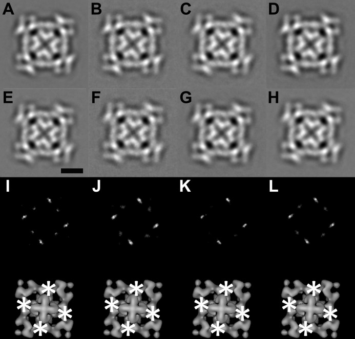
FIGURE 1.
Two-dimensional analysis of CaMWT-RyR1 and CaM1234-RyR1 in 4-fold view. A and B, 4-fold symmetrized two-dimensional averages of apo-CaMWT-RyR1 and CaM1234-RyR1, respectively, in low calcium buffer (2 mm EGTA). C and D, 4-fold symmetrized two-dimensional averages of Ca2+-CaMWT-RyR1 and CaM1234-RyR1, respectively, in high calcium buffer (0.1 mm CaCl2). E and F, 4-fold symmetrized two-dimensional averages of the RyR1 control in low calcium buffer. G and H, 4-fold symmetrized two-dimensional averages of the RyR1 control in high calcium buffer. Scale bar = 10 nm (E). I–L, upper panels, difference maps generated by subtracting images in E–H from A–D, respectively. The white regions in I–L are the positions of positive differences. The biggest differences are in bright white. Lower panels, the positive differences (asterisks) in the upper panels superimposed on the bottom view of a three-dimensional reconstruction of RyR1.

