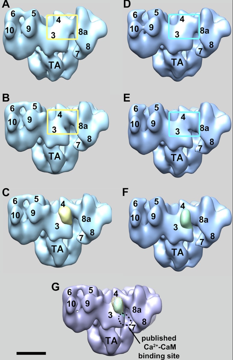
FIGURE 2.
Three-dimensional localization of RyR1-bound CaM1234 at low and high calcium. A and B, solid-body representations of the three-dimensional reconstructions of CaM1234-RyR1 complexes and the CaM-free RyR1 control, respectively, in low calcium buffer. The yellow boxed regions highlight the areas with the biggest difference. C, subtraction of B from A yields four symmetrically related positive differences, one of which is shown superimposed on the RyR1 control (yellow mass) and which is attributed to RyR1-bound CaM1234. D and E, solid-body representations of the three-dimensional reconstructions of CaM1234-RyR1 complexes and the CaM-free RyR1 control, respectively, in high calcium buffer. The cyan boxed regions highlight the areas with the biggest difference. F, the green mass is the result of subtraction of E from D and represents RyR1-bound CaM1234 in high calcium buffer. G, locations of RyR1-bound CaM1234 in low (yellow mesh) and high (green solid) calcium buffers. Both were localized on domain 3 close to domain 4, which closely matches the previously published apo-CaMWT site. The numerals on the cytoplasmic assembly indicate the distinguish domains as numbered previously (30). TA, transmembrane assembly. Scale bar = 10 nm.

