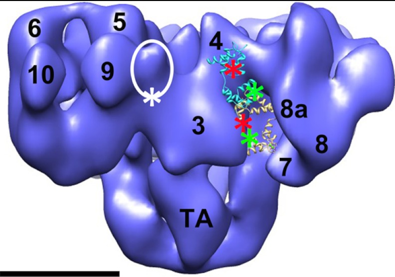
FIGURE 5.
Hypothetical model of FRET pair locations on RyR1-bound apo-CaM, Ca2+-CaM, and FKBP12.6. The white circled region indicates the location of FKBP12.6 on the RyR1 surface, which is between domains 3 and 9. The white asterisk shows one possible donor fluorophore location on the FK506-binding protein. The red asterisks are the potential acceptor fluorophore locations on the N-terminal lobe of apo-CaM (backbone in cyan) and Ca2+-CaM (backbone in yellow). The green asterisks are the acceptor locations on the C-terminal lobe. CaMs were positioned and oriented so as to be consistent with both the cryo-EM localizations and the FRET data that indicate small differences between donor/acceptor fluorophore pairs in apo-CaM versus Ca2+-CaM. Scale bar = 10 nm. The Protein Data Bank code for apo-CaM is 2IX7 (35), and that for Ca2+-CaM is 2BCX (32).

