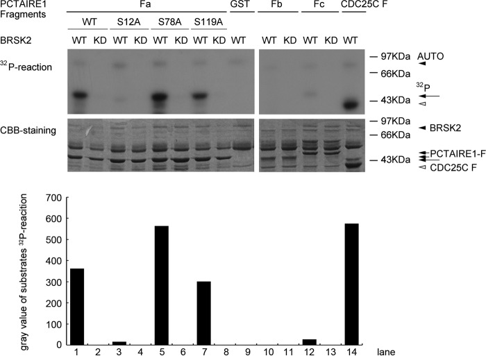
FIGURE 3.
In vitro phosphorylation performance of BRSK2 (wide-type WT or kinase-dead mutant KD) phosphorylation on PCTAIRE1. HA-BRSK2 was expressed in 293T for kinase activity assay. PCTAIRE1 deletion mutants (Fa, Fb, and Fc fragments described in Fig. 2C) and site-directed mutants of Fa (S12A, S78A, and S119A) were used as BRSK2 substrates. The arrowheads in 32P-reaction visualized by autoradiography (top panel) indicated the bands of BRSK2 auto-phosphorylation, PCTAIRE1 fragments phosphorylation and positive control CDC25C F phosphorylation by BRSK2. Corresponding bands using CBB (Coomassie Brilliant Blue) staining (bottom panel) were also arrow-pointed. GST protein showing no BRSK2 phosphorylation signal was used as negative control.

