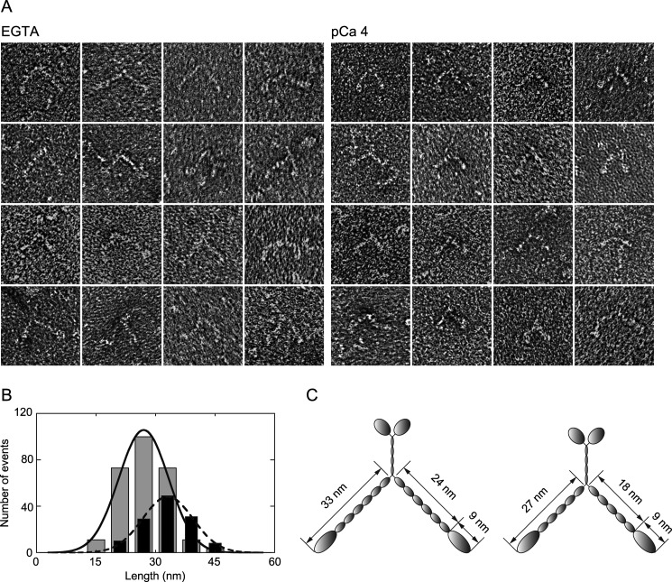
FIGURE 3.
Myosin XI morphology. A, electron micrographs showing morphology of negatively stained myosin XI in EGTA (left panels) and at pCa 4 (right panels). B, lengths from the tip of the head to the base of the neck in EGTA (black bars) and at pCa 4 (gray bars). The means of fits were 32.8 and 27.1 nm, and the numbers of measurements were 128 and 272, respectively. C, illustration of myosin XI before (left) and after (right) the dissociation of CaMs.

