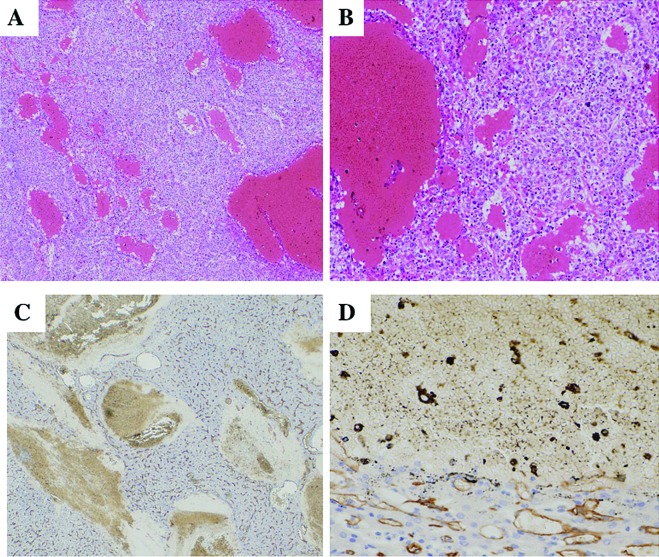
Figure 3.
Histological features of peliotic changes. (A and B) Small and large blood lakes are observed in moderately differentiated HCC and irregular dilatation of sinusoid-like blood spaces of the tumor (H&E stain). (C and D) Immunostaining for CD34 shows no positive cells along the spaces of peliotic change.

