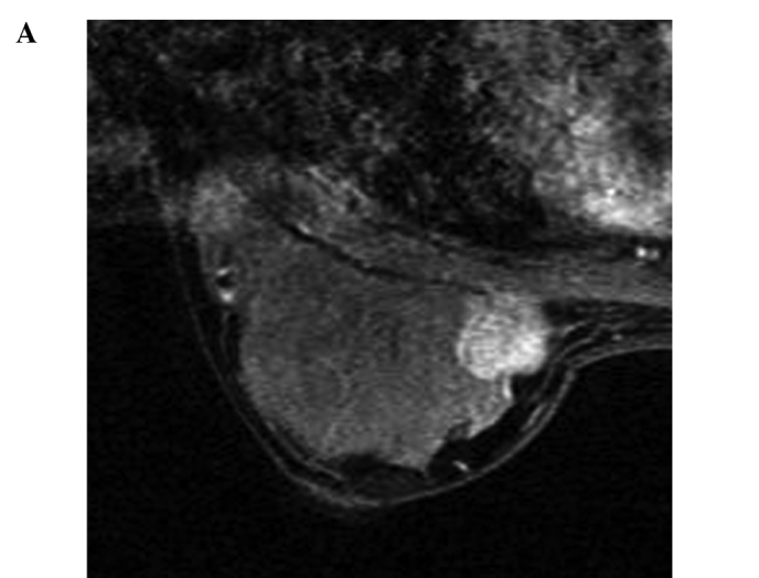
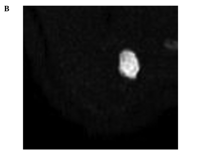
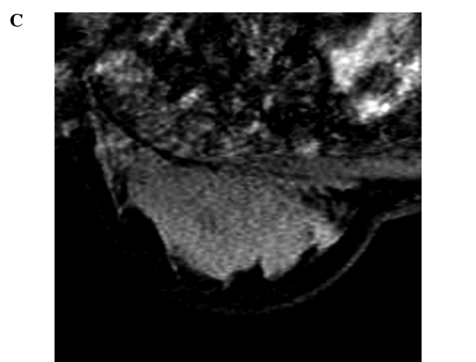
Figure 2.


(A) A 26-year-old woman with invasive ductal carcinoma of the right breast. Three-dimensional contrast-enhanced fat-suppressed MRI of the breast acquired before NAC reveals a localized enhancing mass. (B) Before NAC, the tumor is clearly detected as an area of high intensity on the diffusion-weighted image. (C) Three-dimensional contrast-enhanced fat-suppressed MRI of the breast acquired after NAC reveals a significantly decreased residual mass. (D) After NAC, the tumor is detected as an area of high intensity on the diffusion-weighted image, smaller in size compared to that on the initial MRI. The change in ADC is 75.2% and the response rate is 91%. The extent of the residual tumor on the enhanced MRI corresponded approximately to the surgical findings by mastectomy.
