FIGURE 2.
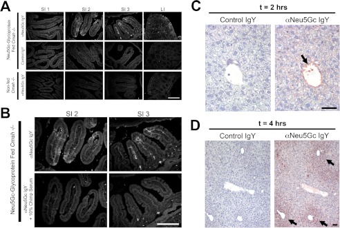
Intestinal uptake of Neu5Gc seen only in Neu5Gc-glycoprotein-fed mice. A, frozen sections of indicated intestine from Neu5Gc-glycoprotein-fed mice 2 h after feeding. Sections are stained with 1:5000 αNeu5Gc IgY (top row) showing prominent uptake (white arrows) in the middle (SI2) and terminal (SI3) part of the small intestine, agreeing with Fig. 1D. Control IgY shows absence of staining on same tissues (middle row). Importantly, tissues from non-fed Cmah−/− mice show absence of staining with αNeu5Gc IgY (bottom row). B, to control for the staining in A, we repeated staining in SI2 and SI3 with and without 10% chimpanzee serum during the primary antibody incubation step. Chimpanzee serum is a rich source of Neu5Gc-containing glycoproteins and blocked staining seen by αNeu5Gc IgY in the SI segments (bottom row). C and D, uptake of Neu5Gc in Neu5Gc-glycoprotein-fed mice by the liver. To determine whether Neu5Gc derived from Neu5Gc-glycoprotein feeding is trafficked to peripheral tissues, we harvested livers of fed mice at 2 h (C) and 4 h (D). Immunohistochemistry using αNeu5Gc IgY revealed staining (black arrows) in the portal vein at 2 h (C), periportal hepatocytes at 4 h (D), and increasingly diffuse periportal hepatocytes at 6 h (figure not shown). Scale bar, 100 μm.

