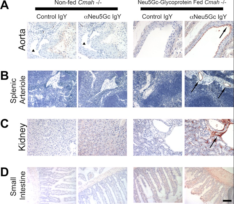
FIGURE 4.
Long term Neu5Gc-glycoprotein feeding leads to metabolic incorporation of Neu5Gc with a human-like tissue distribution I. A–D, paraffin-embedded sections of aorta (A), spleen (B), kidney (C), and small intestine (D) of 3-week Neu5Gc-glycoprotein-fed mice were stained with 1:5000 control IgY or αNeu5Gc IgY (two right columns). Brown staining (see black arrows) with αNeu5Gc IgY, which is not seen with Control IgY, indicates that Neu5Gc is incorporated into these tissues. Similar sections from non-fed Cmah−/− tissues were also stained with control IgY or αNeu5Gc IgY (two left columns), which showed no staining. Collapsed lumen of non-fed aorta (A, left columns) is marked (see black arrowhead). Scale bar, 100 μm.

