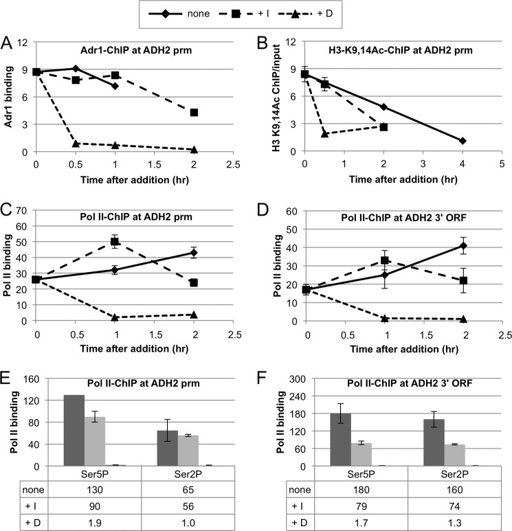
FIGURE 4.
Continuous Snf1 activity is not needed for Adr1 and RNA pol II binding, histone H3-K9,14 acetylation, or RNA pol II CTD phosphorylation. A, Adr1-Myc binding after inhibiting Snf1as and adding glucose. Adr1-Myc binding in strain TYY1077 was measured by ChIP after derepression as described in the legend to Fig. 2, but the cultures were derepressed for only 2 h in low glucose medium. The final concentration of 2NM-PP1 was 10 μm. Duplicate 60 samples were removed at the times indicated. A 10-ml portion of this sample was used for RNA preparation and analysis. The remaining 50 ml was used for ChIP analysis for Adr1-Myc13 as described under “Experimental Procedures.” ChIP-grade monoclonal antibody 9E10 from Santa Cruz Biotechnology (anti-Myc) was used. The data are expressed as binding (ChIP/input) for ADH2prm relative to ChIP/input at the TEL region used as a reference. B, histone H3-K9,14 acetylation after inhibiting Snf1as or adding glucose. Strain TYY1077 was grown and treated as described in the Fig. 2 legend. ChIP for K9,14-acetylated histone H3 was performed using anti-histone H3-K9,14 antisera from Santa Cruz Biotechnology as described under “Experimental Procedures.” The data are expressed as the ratio of ChIP/input without normalizing to the level of acetylation at the TEL region that changed less than 2-fold with any of the treatments. C and D, RNA pol II remains bound to the ADH2 transcription start sites (tss) and ORFs after inhibiting Snf1as. Growth and treatment of the cells is described in the Fig. 2 legend. RNA pol II binding was measured by ChIP as described under “Experimental Procedures” using ChIP-grade Abcam anti-pol II antibody 8WG16. The data are expressed as binding (ChIP/input) for ADH2tss and POX1tss relative to ChIP/input at the TEL region used as a reference. E and F, RNA pol II CTD associated with ADH2 is phosphorylated on Ser-2 and Ser-5 after Snf1as inhibition. The cultures for ChIP analysis were derepressed for 6 h (none), and 2NM-PP1 (+I) or glucose (+D) was added after 2 h of derepression. Abcam anti-pSer-5 and anti-pSer-2 polyclonal antisera were used as described under “Experimental Procedures.” The data are expressed as binding (ChIP/input) for ADH2tss and ORF relative to ChIP/input at the TEL region used as a reference.

