FIGURE 5.
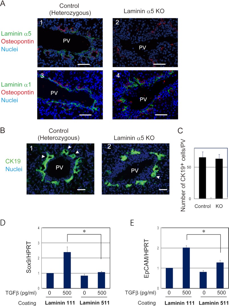
Laminin α5 is dispensable for the lineage determination of hepatoblasts to cholangiocytes. A, laminin α5 is not observed at the basal side of cholangiocytes in laminin α5 KO livers, whereas laminin α1 is normally expressed. The control and laminin α5 KO livers were stained with anti-laminin α5 (panels 1 and 2) or anti-laminin α1 (panels 3 and 4) and anti-osteopontin antibodies. Bars represent 50 μm. B, CK19+ cholangiocytes are observed around the portal vein in laminin α5 KO liver. E17.5 liver sections were stained with anti-CK19 antibody. Arrowheads indicate luminal structures surrounded by CK19+ cells. C, numbers of CK19+ cholangiocytes in the control and mutant livers. The number of CK19+ cholangiocytes around the portal vein was counted in liver sections prepared from two control and three mutant livers. There was no statistically significant difference between the control and KO. A t test was performed by Microsoft Excel software. D and E, TGFβ induces Sox9 and EpCAM, cholangiocyte markers, in hepatoblasts more efficiently on α1-containing laminin than on α5-containing laminin. Hepatoblasts were isolated from E14.5 liver and cultured on dishes coated with α1-containing laminin (laminin 111) or α5-containing laminin (laminin 511). At day 3 of culture, cells were stimulated with 500 pg/ml TGFβ for 24 h. Gene expression was examined by quantitative PCR. Cultures were repeated three times independently. A t test was performed by Microsoft Excel software. *, p < 0.05. HPRT, hypoxanthine phosphoribosyltransferase; PV, portal vein. Error bars in panels C, D and E represent standard deviation.

