FIGURE 6.
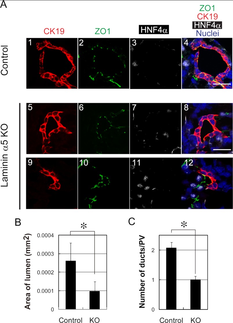
Defects of bile duct formation in laminin α5 KO liver. A, duct structures in the control and laminin α5 KO livers are characterized by the expression of CK19, ZO1, and HNF4α. All three duct structures are associated with a single lumen surrounded by cells with tight junctions recognized by ZO1 staining (green). Ducts in panels 1–8 are totally surrounded by CK19+HNF4α− cholangiocytes. On the other hand, an immature duct in panels 9–12 is surrounded by CK19+HNF4α− cholangiocytes and CK19−HNF4α+ hepatoblasts. B, the lumen size of the bile duct is reduced in laminin α5 KO liver. The lumen surrounded with CK19+ cholangiocytes was selected, and its area was measured by ImageJ. A t test was performed by Microsoft Excel software. *, p < 0.05. C, the number of duct structures is reduced in laminin α5 KO liver. CK19+ duct structures around the portal vein were counted in more than 10 sections in each liver tissue. More than 50 portal areas were examined for each embryonic liver. A t test was performed by Microsoft Excel software. *, p < 0.05. Scale bars represent 20 μm. Error bars represent standard deviation.

