
FIGURE 1.
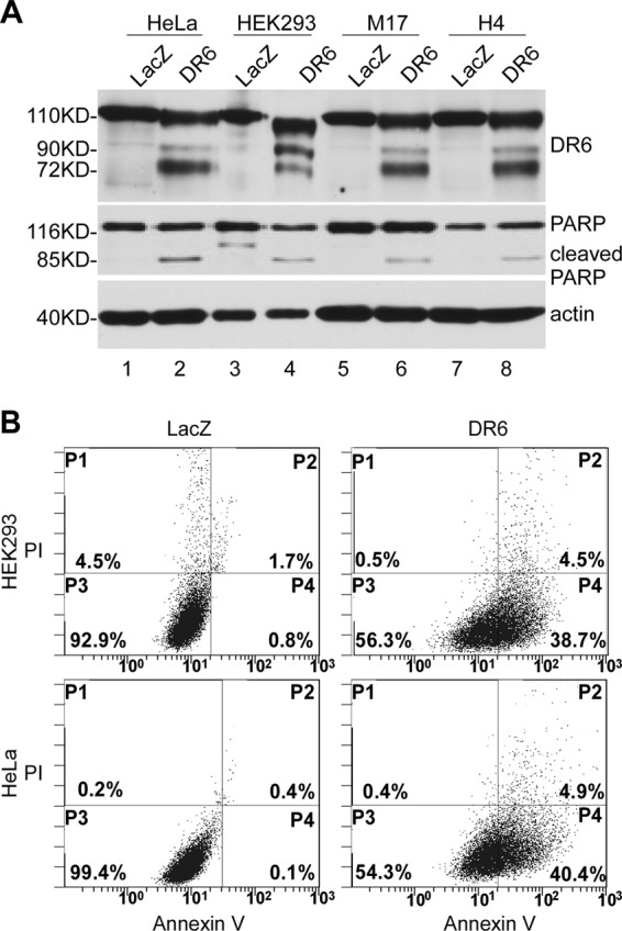
Overexpression of DR6-induced apoptosis in various types of cells. A, cells were transiently transfected with either the DR6-expressing construct pcDNA3.1/Myc-His/DR6 (DR6), which expresses a DR6-Myc fusion protein; or a control plasmid, pcDNA3.1/Myc-His/LacZ (LacZ), which expresses Myc-tagged LacZ protein. 24 h after transfection, cells were harvested, and the lysates were separated by SDS-PAGE, followed by Western blot analysis. Expression of DR6 and LacZ was detected using anti-Myc antibody (top panel). LacZ was detected as a 116-kDa peptide as expected (lanes 1, 3, 5, and 7). DR6 was detected as several bands within the molecular mass range 72–110 kDa, resulting from different glycosylation (14). PARP cleavage was detected in the DR6-expressing samples (lanes 2, 4, 6 and 8, middle panel) but not in the LacZ-transfected cells (lanes 1, 3, 5, and 7). This membrane was also reprobed with anti-actin antibody to indicate relative loading of samples (bottom panel). B, representative FACS scatter plots of three independent experiments are shown. DR6- (right column) or LacZ- (left column) transfected HeLa (bottom row) and HEK293 cells (top row) were double stained with Annexin-V-enhanced green fluorescent protein and PI. Fluorescence was detected using a fluorescence-activated Epics XL-MCL Flow Cytometer to analyze necrotic (PI-positive, quadrant P1), nonapoptotic (negative for both dyes, quadrant P3), early apoptotic (Annexin-positive/PI-negative, quadrant P4), and late apoptotic cells (positive for dyes, quadrant P2).