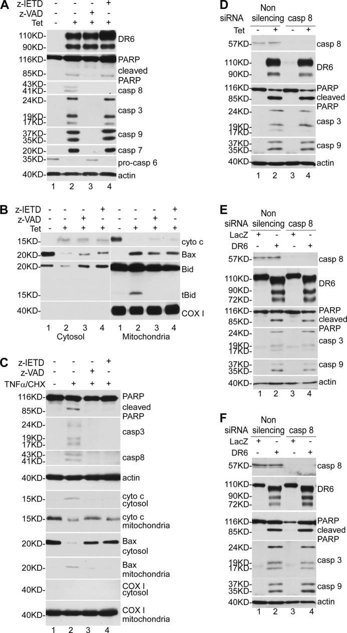
FIGURE 3.

Caspase-8 is not required for DR6-induced apoptosis. A, T-RE HeLa-inducible cells were cultured in the presence or absence of caspase inhibitors and induced for DR6 expression for 48 h. Half of the cells were directly lysed and subjected to Western blot analysis, and the other half were used for preparation of cytosolic extracts and mitochondria-containing membrane fraction. DR6 expression, PARP cleavage, and caspase activation were determined as described in the legend to Fig. 2. Because the active form of caspase-6 is hardly detectable, caspase-6 activation was determined by the decrease in procaspase-6. The general caspase inhibitor Z-VAD was used at 100 μm (lane 3), and the caspase-8-specific inhibitor Z-IETD was used at 100 μm (lane 4). In lanes 1 and 2, cells were treated with vehicle only. The bottom panel is the reprobe of the membrane of the fifth panel with anti-actin antibody to indicate relative loading of samples. B, neither Z-VAD nor Z-IETD blocked cytochrome c release and Bax translocation induced by DR6. Cellular fractionation and Western blot analysis were performed as described under “Experimental Procedures.” The mitochondrial protein COX I was detected only in the membrane fraction (bottom panel), indicating that the mitochondria remained intact during the sample preparation. As shown by previous studies, in addition to tBid, a significant Bid proprotein was detected in the mitochondrial fraction (19, 20). C, caspase-8 inhibitor abrogates apoptosis induced by co-treatment with TNFα and CHX. Cells were pretreated with caspase inhibitors or vehicle only for 30 min and induced for apoptosis by TNFα and CHX for 6 h. PARP cleavage, caspase activation, cytochrome c release, and Bax translocation were determined as described by Western blot analysis. The fourth panel is the reprobe of the membrane of the third panel with anti-actin antibody to indicate relative loading of samples. The mitochondrial protein COX I was detected only in the membrane fraction (bottom panel), indicating that the mitochondria remained intact during sample preparation. Knockdown of caspase-8 had no effect on DR6-induced apoptosis in HeLa cells (D), HEK293 cells (E), and H4 cells (F). Top panel shows Western blot for caspase-8; second panel, Western blot for DR6; third panel, Western blot for PARP; fourth panel, Western blot for caspase-3; fifth panel, Western blot for caspase-9; bottom panel, membranes in the fifth panel were reprobed with anti-actin antibody to indicate relative loading of samples.
