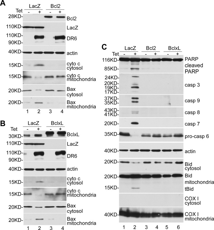
FIGURE 4.
Overexpression of antiapoptotic protein Bcl-2 and Bcl-xL inhibited DR6-induced apoptosis. Cell lysis and fractionation were performed as described previously. A and B, after 24-h transfection, a significant amount of Bcl-2 and Bcl-xL was detected (lanes 3 and 4, top panel). Because the recombinant Bcl-2 and Bcl-xL were expressed as a FLAG-tagged protein, the recombinant Bcl-2 and Bcl-xL were detected with a slower migration rate than endogenous Bcl-2 and Bcl-xL (compare lanes 3 and 4 with lanes 1 and 2). This membrane was also reprobed with anti-actin antibody to indicate relative loading of samples (fourth panel). Second and third panels show tetracycline (Tet)-induced DR6 expression, respectively; fifth and sixth panels, cytochrome c release from mitochondria; seventh and eighth panels, Bax translocation from cytosol to mitochondria. C, top panel shows Western blot for PARP; second to sixth panels, Western blots for caspases; seventh panel, Western blot for actin to indicate relative loading of lysate samples; eighth and ninth panels, Western blots for Bid; tenth and eleventh panels were stained for mitochondrial protein COX I.

