
FIGURE 6.
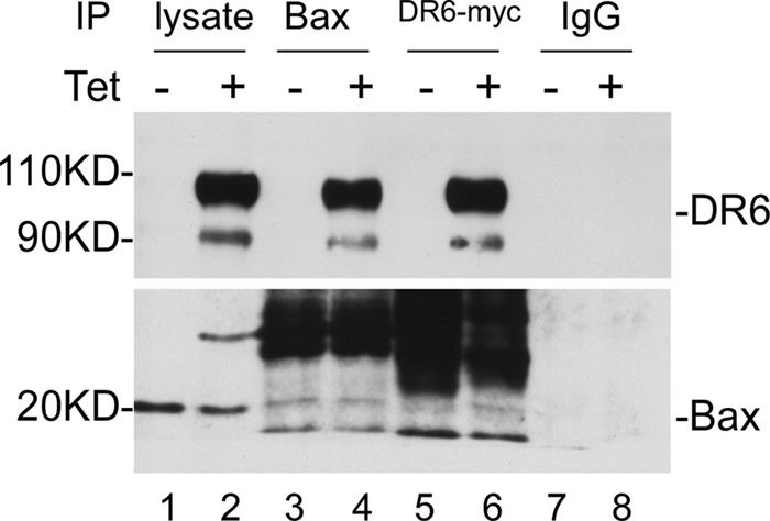
Co-immunoprecipitation of DR6 with Bax. 48 h after induction of DR6 expression, total cell lysates were immunoprecipitated with anti-Bax (lane 4) and with anti-Myc (lane 6) or with a control rabbit IgG (lane 8) and then probed with anti-DR6 (upper panel) and anti-Bax (lower panel). The endogenous DR6 (lane 1, top panel) is hardly visible without prolonged exposure.