FIGURE 2.
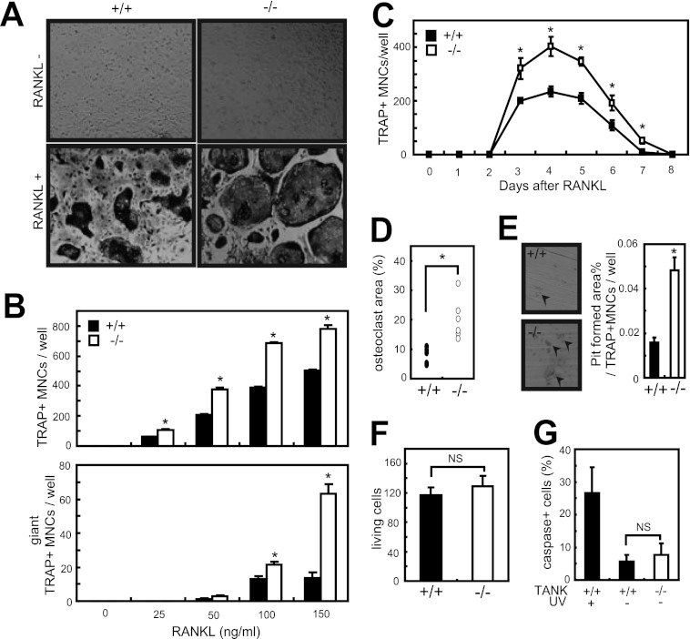
Osteoclast differentiation is increased in Tank−/− MDMs. A, MDMs from wild-type and Tank−/− mice were cultured in the presence of 100 ng/ml RANKL. After 3 days, TRAP staining was performed. B, numbers of TRAP-positive multinucleated (>3 nuclei/cell identified in a ×10 field) cells and giant TRAP-positive multinucleated (>20 nuclei/cell) cells were counted. Error bars: S.E. (n = 3). *, p < 0.05 versus wild-type. C, sequential TRAP staining to count the numbers of osteoclasts from wild-type and Tank−/− MDMs (RANKL, 50 ng/ml). Error bars: S.E. (n = 3). *, p < 0.05 versus 0 h. D, osteoclast areas (percentages of TRAP-positive multinucleated cells relative to the total area) were measured using ImageJ (RANKL, 100 ng/ml) and normalized by osteoclast number. Error bars: S.E. (n = 6). *, p < 0.05. E, formation of resorption pits by osteoclasts induced from wild-type or Tank−/− mice (RANKL, 50 ng/ml). Error bars: S.E. (n = 3). *, p < 0.05. F, MDMs were stained with trypan blue, and the numbers of viable cells were determined under a light microscope. G, apoptosis of wild-type and Tank−/− MDMs. Caspases were stained using a Poly Caspase Assay Kit, and the numbers of caspase-positive cells were counted under a fluorescence microscope. The data shown are representative of three (A, B, D, F, and G) and two (C and E) independent experiments.

