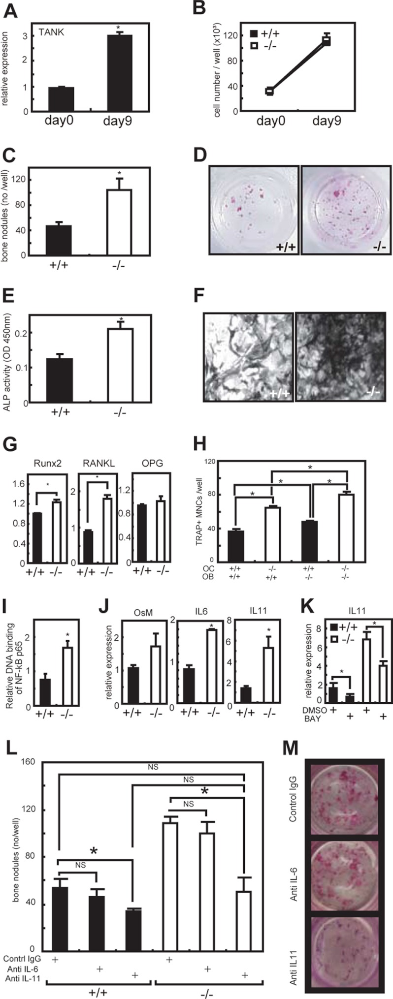
FIGURE 6.
Increased bone formation by Tank−/− calvarial osteoblasts. A, calvariae from wild-type mice were treated with an osteoblast-inducing reagent for 9 days. The mRNA levels of TANK were measured by quantitative PCR. Error bars: S.E. (n = 3). *, p < 0.05 versus d 0. B, numbers of osteoblasts in culture. C, osteoblasts (after a 9-day culture of calvariae) were stained with a calcified nodule staining kit, and the numbers of bone nodules were counted. Error bars: S.E. (n = 3). *, p < 0.05 versus wild-type. D, representative images of calcified nodules in C. E, ALP activities in homogenates of the osteoblasts. Error bars: S.E. (n = 3). *, p < 0.05 versus wild-type. F, representative images of ALP staining in osteoblasts. G, mRNA levels of Runx2, RANKL, and OPG were measured by quantitative PCR. Error bars: S.E. (n = 3). *, p < 0.05, versus wild-type. H, bone marrow cells (OC) and calvariae (OB) were cocultured in the presence of 1α,25(OH)2D3 for 10 days. The numbers of TRAP-positive multinucleated cells were counted. Error bars: S.E. (n = 3). *, p < 0.05. I, DNA binding activity of NF-κB p65 in osteoblasts (after a 9-day culture of calvariae) was measured using a TransAM Transcription Factor Assay Kit. Error bars: S.E. (n = 3). *, p < 0.05. J, quantitative PCR analyses OncostatinM (OsM), IL-6, and IL-11 in osteobalsts. Error bars: S.E. (n = 3). *, p < 0.05. K, 5 mm NF-κB inhibitor BAY117085 was added to the osteoblast culture for 24 h and IL-11 mRNA level was quantified. Error bars: S.E. (n = 3). *, p < 0.05. L, calvarial osteoblasts were cultured with neutralizing antibodies of IL-6 and IL-11. After a 9-day culture, cells were stained with a calcified nodule staining kit, and the numbers of bone nodules were counted. Error bars: S.E. (n = 3). *, p < 0.05. M, representative images of bone nodules formed by Tank−/− calvarial osteoblasts in L.

