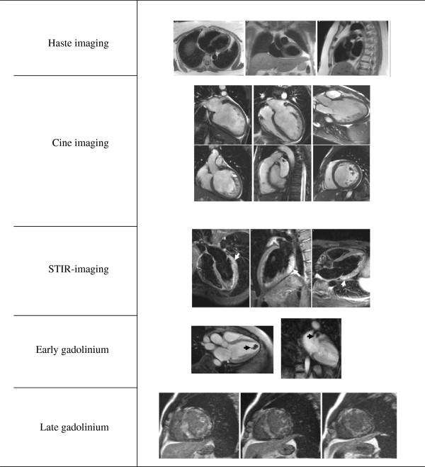
Figure 1.
Standard cardiomyopathy protocol. Standard cardiomyopathy protocol including HASTE, cine imaging, T2-weighted-imaging (STIR) searching for inflammation/edema (white arrows), early gadolinium imaging to detect thrombus (black arrow) and LGE to identify fibrosis or infiltration (diffuse LGE typical of amyloidosis).

