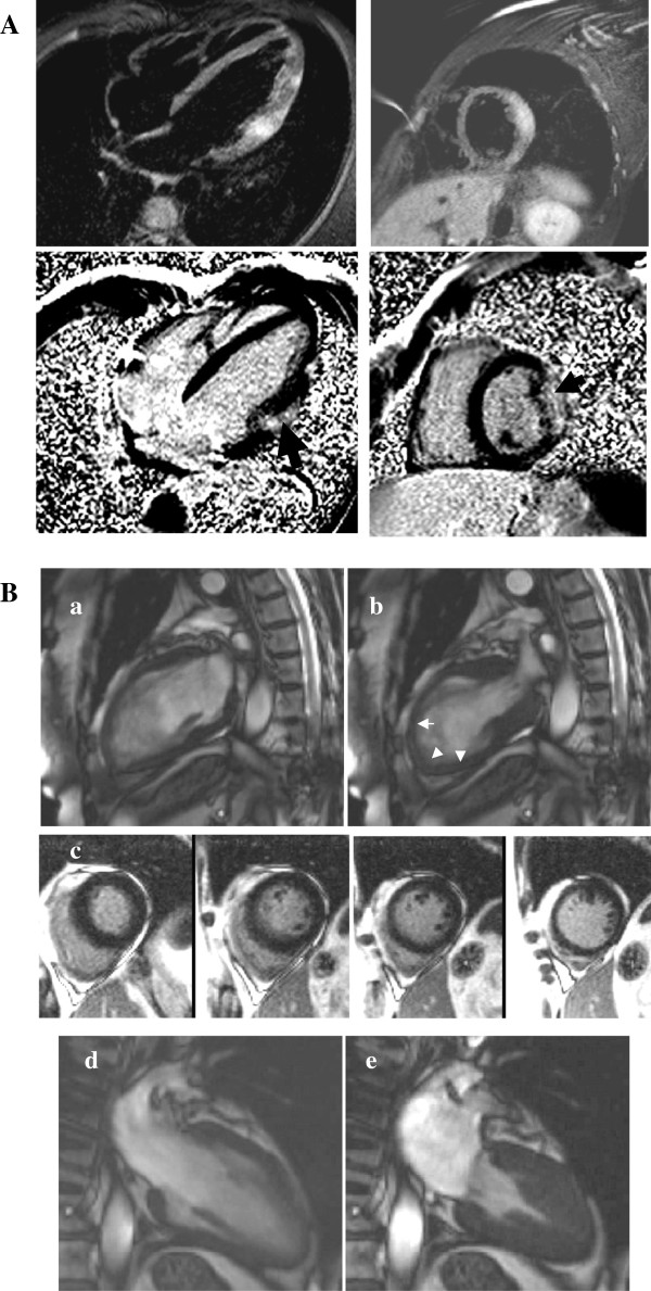
Figure 7.
Acute chest pain syndromes (A) Acute viral myocarditis displaying edema on STIR images (a-b, arrows) and typical sub-epicardial LGE (c-d, arrows). (B) Tako-tsubo with acute apical ballooning of the LV (a: diastole, b: systole displaying apical akinesia, arrows) without LGE (c) followed by complete LV functional recovery at follow-up (d: diastole, e: systole).

