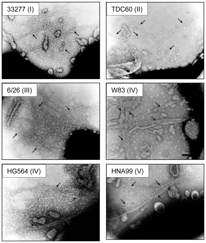
Figure 3. Transmission electron microscopic observation of FimA fimbriae on the bacterial cell surface.
P. gingivalis ATCC 33277 Δmfa1 Δfim cluster cells with fimA from 33277, TDC60, 6/26, W83, HG564 and HNA99 introduced by using an expression vector. Samples were negatively stained with 1% ammonium molybdate. Arrows indicate fimbrial structure. Some fimbriae appear to be bundled. Bars show 0.2 µm.

