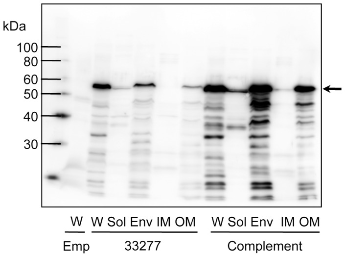
Figure 4. Immunoblot analysis for PgmA using whole-cell sonicates.

Whole-cell sonicates (W) were fractionated into soluble (Sol), envelope (Env), inner membrane (IM) and outer membrane (OM) fractions. Samples were denatured in an SDS-containing buffer with 2-mercaptoethanol by heating at 100°C for 10 min, then subjected to SDS-PAGE and immunoblot analysis. Emp denotes 33277 Δmfa1 Δfim cluster/pT-COW::ragAP, carrying empty vector, used as a negative control; 33277 denotes the wild-type strain; Complement denotes 33277 Δmfa1 Δfim cluster carrying pT-COW::ragAP::fimX-pgmA-fimA. An arrow indicates PgmA as a 60-kDa protein. Degradation bands (below the 60-kDa) were also visualized because PgmA was highly sensitive to intrinsic proteases of this bacterium [13].
