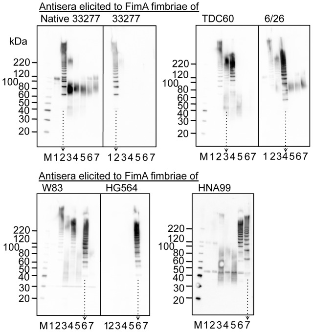
Figure 8. Immunoblot analysis using whole-cell sonicates partially denatured.
Whole-cell sonicates were denatured in an SDS-containing buffer with 2-mercaptoethanol by heating at 70°C for 10 min, and subjected to SDS-PAGE and immunoblot analysis by using antisera, 1,000-fold dilution, from mice immunized with purified FimA fimbriae. Antigen samples were as follows: P. gingivalis ATCC 33277 Δmfa1 Δfim cluster (FimA deficient, lane 1), and the wild-type strains of ATCC 33277 (lane 2), TDC60 (lane 3), 6/26 (lane 4), W83 (lane 5), HG564 (lane 6), and HNA99 (lane 7). M denotes a standard marker. W83 rarely produces FimA protein and fimbriae. Note that ladder bands are specific for FimA fimbriae whereas smear bands between 40–80 kDa are nonspecific. Arrows with dotted lines are placed in order to clearly discriminate each lane.

